SCHEDULE A CONSULTATION
Schedule by phone at (973) 635-5050
or fill out the online consultation request
By submitting this form you agree to be contacted via phone/email/text
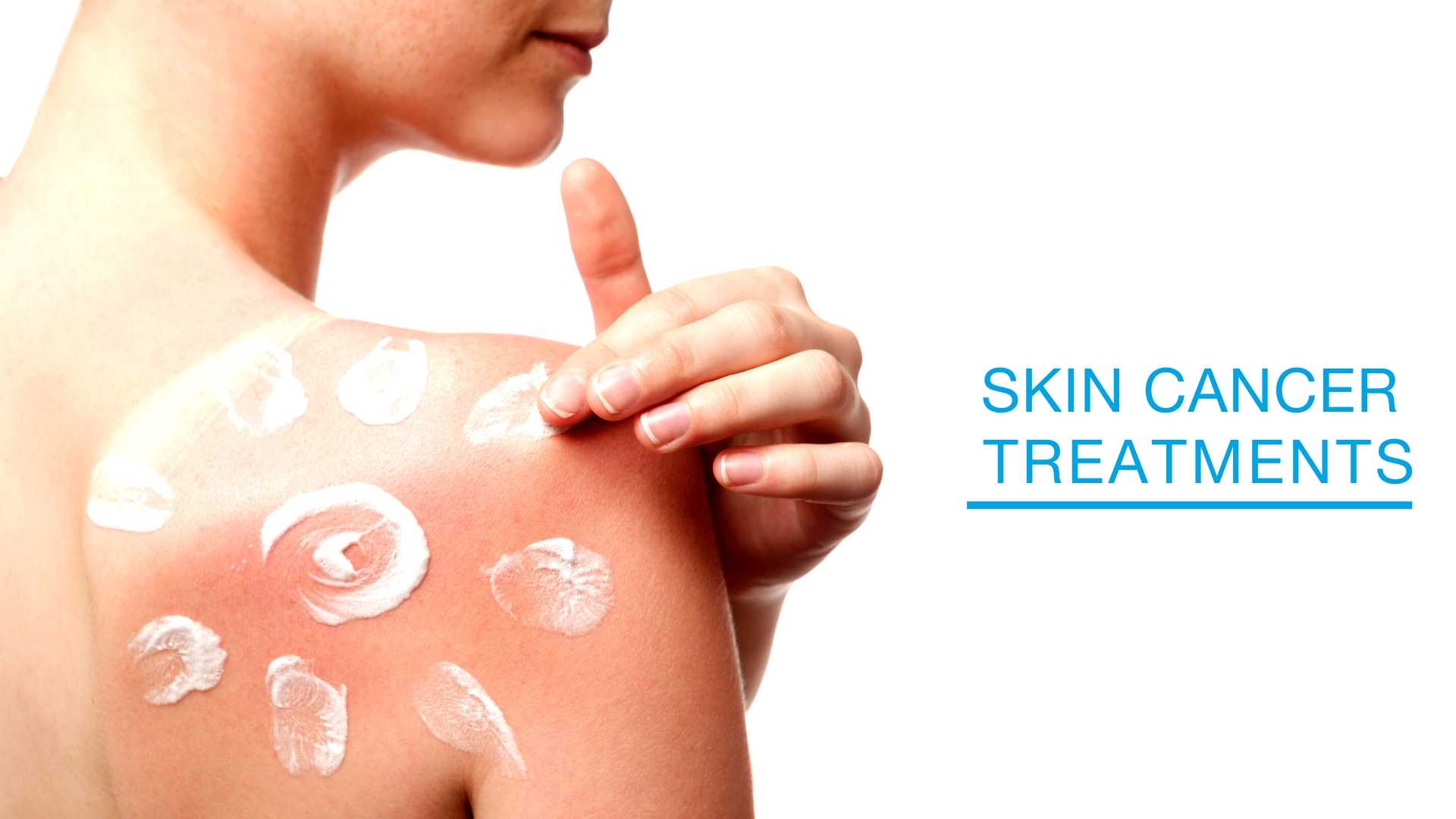
Skin Cancer Treatments at Laser + Skin Institute
What is Skin Cancer?
The underlying cause of skin cancer in older people is often the accumulated damage of many years of excessive exposure to the sun.
In some cases there may be a genetic predisposition to skin cancer – either “cancer in the family” or the inheritance of a type of skin that increases risk for skin cancer. All skin cancers can be successfully treated if they are discovered and treated early. All are potentially disfiguring, and potentially fatal if they metastasize (spread) to other parts of the body.
The three most common forms of skin cancer are:
Basal Cell Carcinoma
Squamous Cell Carcinoma
Melanoma
What is Basal Cell Carcinoma?
Basal cell carcinoma arises in a layer of skin (basal layer) beneath the skin’s surface. It seldom metastasizes, although it may do so if the cancer invades lymph or blood vessels that can carry cancer cells to distant organs. The major spreading mechanism of basal cell carcinoma is by local invasion of surrounding skin tissue. If left untreated, it may become large and disfiguring.
Risk factors for developing basal cell carcinoma are:
- excessive and chronic sun exposure over many years
- a fair (white) skin complexion, especially when hair is blond or red
While basal cell carcinoma has traditionally been a cancer associated with older people, it is now seen in more young adults than in the past.
Early detection of basal cell carcinoma can lead to early treatment and prevention of disfigurement. The most likely places for basal cell carcinoma to develop are areas exposed to sun—face, scalp, ears, neck, shoulders and back.
Criteria to look for in self-examination:
- a small, pearly nodule, which may or may not have telangiectasia (small enlarged blood vessels) on the surface; the nodule increases in size slowly and may form an ulceration in its center; there may be some pigmentation
- a solitary, flat or slightly depressed lesion that is hard to the touch; it may be yellowish or whitish and have indistinct borders
- one or more reddish, scaling plaques that slowly enlarge; these lesions may resemble dermatitis or psoriasis
Any suspicious lesion should be examined immediately by one of our physicians and biopsied if deemed necessary to determine proper treatment.
Early, effective treatment of basal cell carcinoma by a dermatologic surgeon has a cure rate of more than 95%. However, new basal cell carcinomas can develop after treatment, so continued self-examination and regular examination by a dermatologist are important.
When basal cell carcinoma is discovered early and the diagnosis confirmed by biopsy, treatment may be carried out in the dermatologist’s office or an outpatient setting.
Treatment procedures include:
- Curettage: A scalpel is used to scrape away malignant tissue. Electrocautery may be used after curettage to “mop up” any remaining cancer cells. Curettage is used chiefly for superficial carcinoma not previously treated.
- Cryosurgery: Liquid nitrogen is applied to the lesion to destroy malignant tissue by ultra-cold freezing.
- Topical chemotherapy: Cancer cells are destroyed by pharmacologic agents applied to the surface of the skin.
- Surgical excision: The cancer is surgically removed and the skin closed with stitches. This technique is used when the carcinoma is in deeper tissues.
- Mohs microscopic surgery: Surgical removal is performed under a microscope. In this technique, the surgeon can perform surgery layer by layer into the skin, under direct microscopic observation.
- Laser surgery: Cancerous tissue is destroyed by laser beam.
Our physicians will discuss with you the type of treatment that will be most effective.
What is Squamous Cell Carcinoma?
Squamous cell carcinoma develops in the outer layers of the skin. It is capable of metastasizing to other areas of the body if not treated early. It also spreads locally and may cause significant disfigurement.
Risk factors for developing squamous cell carcinoma are:
- excessive, chronic exposure to sun, over many years
- overexposure or chronic exposure to x-rays
- long-term treatment with immunosuppressive drugs
- white skin, especially with blond or red hair
Criteria to look for in self-examination:
- commonly appears as an ulcerated nodule or superficial erosion with poorly defined margins on the skin or lower lip; the lesion persists and does not heal
- a wart-like growth or plaque
- premalignant forms of squamous cell carcinoma include actinic keratosis, cutaneous horns (hard, fibrous growths), and Bowen’s disease (scaling, inflamed-looking plaques)
Any suspicious lesion should be examined immediately by one of our physicians and biopsied if deemed necessary to determine proper treatment.
When a diagnosis of squamous cell carcinoma is confirmed by biopsy, treatment options are similar to those for basal cell carcinoma.
What is Melanoma?
As with basal cell carcinoma and squamous cell carcinoma, excessive and chronic sun exposure is a major risk factor for melanoma. There also is a tendency for melanoma to “run in the family”, and to be associated with a familial trait of having many moles on the body. Melanoma often arises in a pre-existing mole or pigmented lesion. Early diagnosis and treatment of melanoma is essential. Any person with many moles or a family history of melanoma should be examined regularly by one of our Physicians or Physician Assistant. Every adult should self-examine at regular intervals to detect any early indications of melanoma.
Self-examination is done using the A-B-C-D criteria:
- A = Asymmetry (the left side of the lesion is unlike the right side)
- B = Border Irregularity (the lesion has a scalloped or poorly defined border)
- C = Color Variation (not all parts of the lesion are the same color;
within the lesion may be patches of tan, brown, black, pink, white or blue) - D = Diameter (while melanomas are usually greater than 6mm in diameter when diagnosed, they can be smaller. If you notice a mole different from others, or which changes, itches or bleeds even if it is smaller than 6mm, you should see a dermatologist)
It is worth noting that some melanomas do not conform to the A-B-C-D criteria, so any suspicious mole should be examined by one of our physicians.
What if I suspect that I may have skin cancer?
To schedule an appointment with our Physicians, fill up the form below or call our scheduling staff at 973-635-5050.
SCHEDULE A CONSULTATION
Schedule by phone at (973) 635-5050
or fill out the online consultation request
By submitting this form you agree to be contacted via phone/email/text
SCHEDULE A CONSULTATION
Schedule by phone at (973) 635-5050
or fill out the online consultation request
By submitting this form you agree to be contacted via phone/email/text